During my Master’s studies, I worked on a project combining my interest in photoacoustic imaging and elastography. I collaborated with Mr. François Varray from CREATIS, Lyon, to develop a simulation pipeline for shear wave imaging to assess tissue stiffness, a key indicator in diagnosing conditions like cancer or cardiovascular diseases.
The main objective of the project was to simulate the propagation of shear waves in biological tissues, enabling us to visualize tissue stiffness. We used MATLAB-based tools—k-Wave for wave propagation and MUST for ultrasound signal simulation.
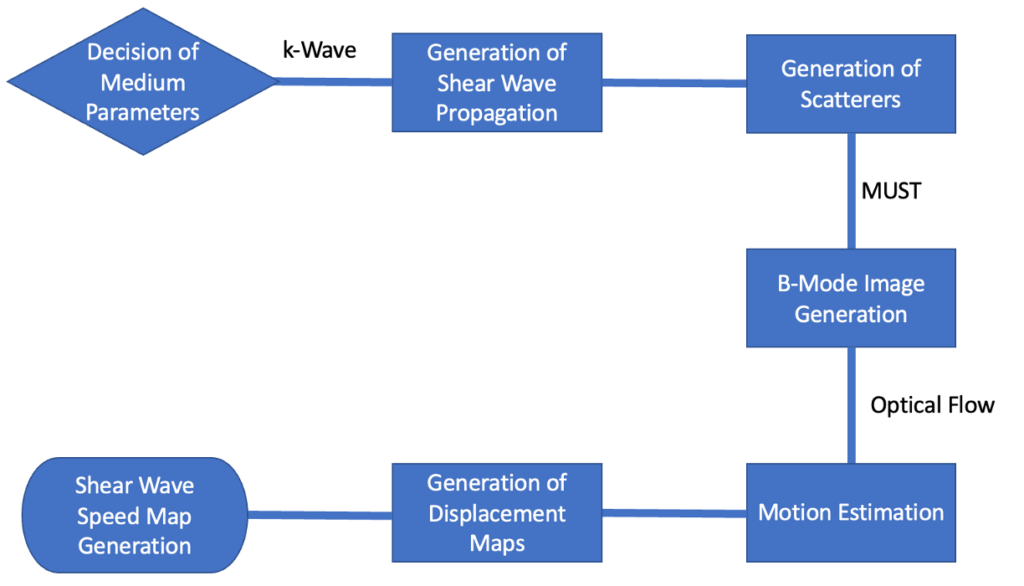
The pipeline consists of following steps
Generation of Shear Waves
Shear waves were simulated using k-Wave, representing wave propagation in both homogeneous and inhomogeneous media. This is akin to how real tissue behaves when subjected to mechanical waves, helping us understand stiffness variations.
Scatterer Generation & B-Mode Imaging
Scatterers were introduced to simulate the tissue structure, and B-Mode images were generated using the MUST toolbox, which is a common ultrasound imaging technique. This allowed us to visualize how shear waves interact with different tissue types.![Scatterer and B-Mode Image]
(Insert scatterer and B-mode image)Motion Detection
By applying optical flow algorithms, we could detect tissue displacement caused by the shear waves. The motion vectors enabled us to reconstruct a displacement map, highlighting areas with varying stiffness.![Displacement Map with Motion Vectors]
(Insert image of displacement map)Shear Wave Speed Map
Although this part was not fully realized during the project, the groundwork for creating shear wave speed maps was laid. These maps will eventually quantify tissue stiffness across the medium.